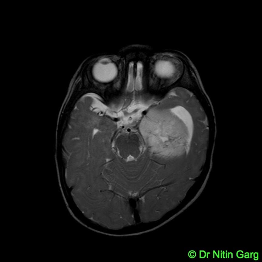
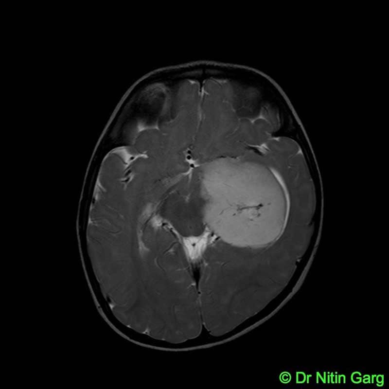
A 9 month old child presented with seizures and irritability. MRI showed a large left medial temporal lesion with significant mass effect (Figure 1). Pre-operatively, possibility of glioma versus peendymoma was considered.
The child underwent left temporal craniotomy, amterior temporal lobectomy and tumor decompression. Intra-operatively, the tumor was soft, suckable, moderately vascular. Medial portion of tumor could not be dissected off the brain stem and was left behind. Child made an eventful recovery. He had mild right hemiparesis which recovered over few days.
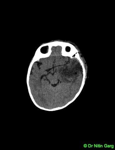
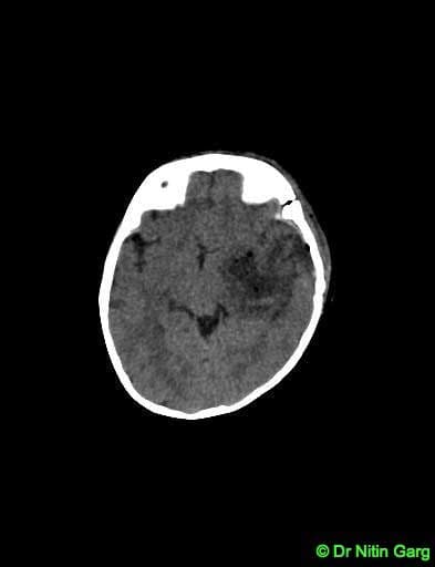
His post-op scan showed adequate tumor decompression and reduction in mass effect. Histopathology was suggesstive of “Ependymoma Grade 2”