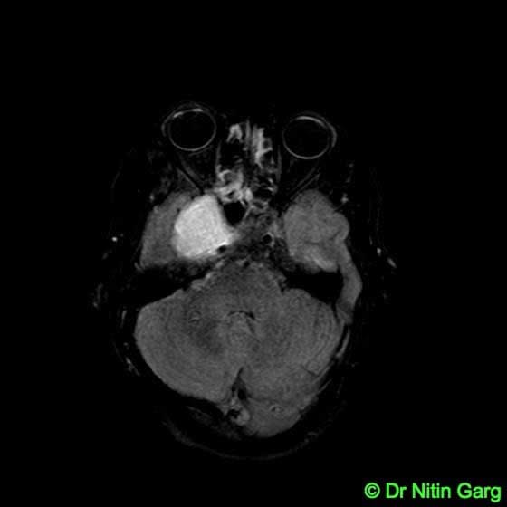
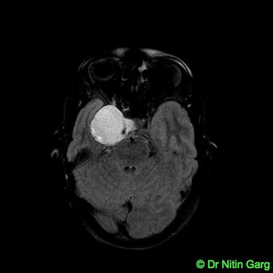
A 25 year old 6 month pregnant lady came with features of right sided facial pain and paralysis of the ocular muscles (cavernous sinus syndrome) of few days duration. MRI of Brain revealed a large right sided temporal skull base lesion with compression of the cavernous sinus. A possibility of hemangioma was considered. In view of significant neurological deficits, she was taken up for surgical removal of the tumor instead of waiting for the completion of pregnancy.
Brain surgery during pregnancy is challenging due to need for antiepileptic drugs, safer anaesthesia, need for repeated scans and risk to the featus due to hemosynamic changes. After discussing with the family regarding the risks, surgery was planned alongwith the anaesthetists, obstetricians and neonatologists. No radiological investigations involving exposure to radiation like CT scan was done.
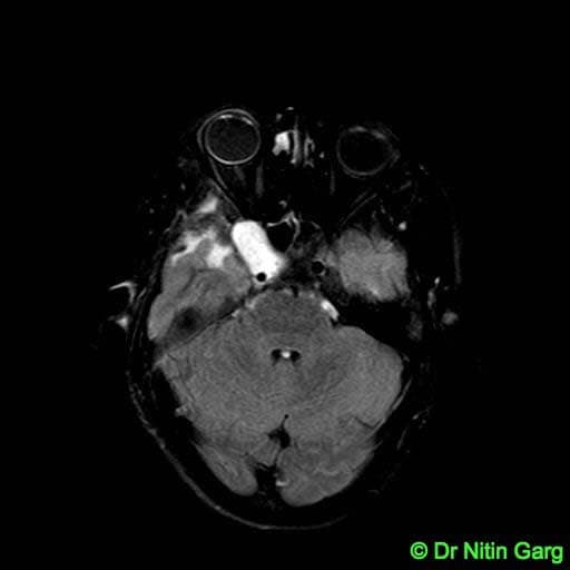
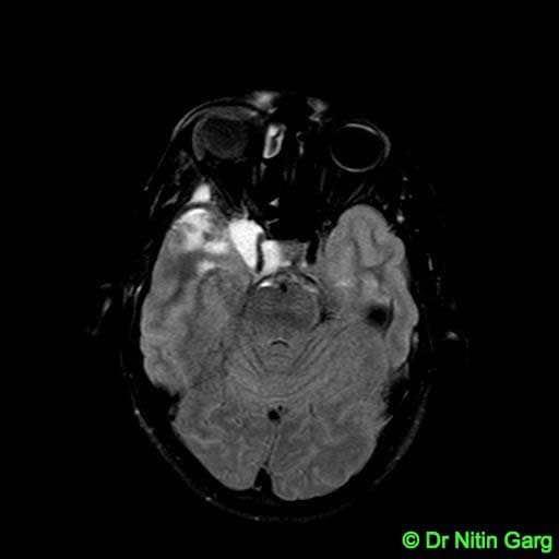
A right temporal craniotomy and zygomatic osteotomy (FTOZ) was done for a better skull base approach and to minimise temporal lobe retraction. Intra-operatively, the tumor was highly vascular with well defined capsule. Tumor excision except the infiltrating portion into the cavernous sinus was excised safely. The patient alongwith the foetus recovered well . A full term child was born subsequently upon completion of gestation. At 6 months follow up, the patient's neurological deficits have recovered. Her MRI scan shows small residual in the cavernous sinus. Gamma Knife Radiosurgery has been planned for the same.
Aids used: Microscope, Neurosurgical drill, Reciprocating saw.