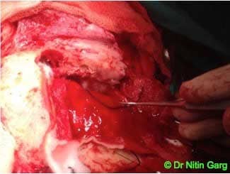
Orbital tumors result in proptosis and visual impairment. Transcranial approaches are utilised for superior, medial, lateral quadrant lesions of the orbit.
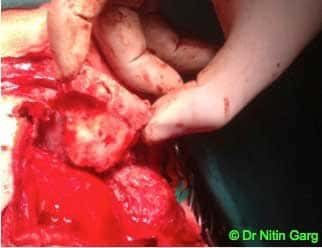
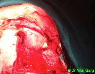
This patient had extra-axial proptosis of the right eye. Imaging was suggestive of extra-conal lesion involving supero-medial quadrant of the orbit. Her vision was intact. She underwent Frontal Craniotomy , orbital osteotomy and gross total excision of the tumor. The lesion was moderately vascular. Following excision, bone was repositioned and fixed with mini-plates.
Post-op, patient recovered well with resolution of proptosis.
Aids used: Neurosurgical drills, Reciprocating saw, Microscope, Navigation, CUSA

.jpg)
.jpg)
.jpg)