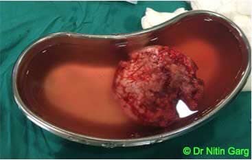
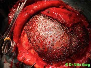
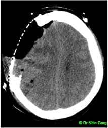
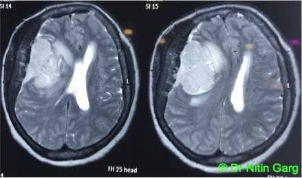
65 year old female patient presented with progressive weakness of left sided limbs and progressive dullness. Her MRI scan showed a large right posterior frontal tumor with significant compression of the underlying brain parenchyma. Associated underlying edema present. Patient underwent navigation assisted craniotomy and Simpson's grade 0 excision of the tumor. The calvarial bone was aslo involved with the tumor and was excised. Dura was infiltrated and excised with adequate margins. Duraplasty was done with autologous pericranial graft. Titanium mesh was used for filling the bony defect.
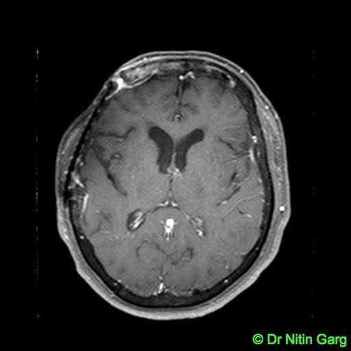
Patient tolerated the procedrue well and recovered uneventfully. Her 3 months follow up MRI shows no residual enhancing tumor.
Aids used: Microscope, Neuronavigation, CUSA, neurosurgical drill

.jpg)