Transcranial endoscopy is very useful for managing deep seated intraventricular tumors and hydrocephalus. Various procedures that can be performed include CSF diversion (endoscopic third ventriculostomy), tumor excision (like colloid cyst) and biopsy of intraventricular tumors (like posterior third ventricular tumors) and paraventricular tumors (as thalamic tumors). 2.7mm scopes are used for transcranial procedures. A 4mm ventricular working port with 4 ports (one for camera and light source, one for irrigation and the third for instruments / bipolar is used for biopsy / tumor resections.
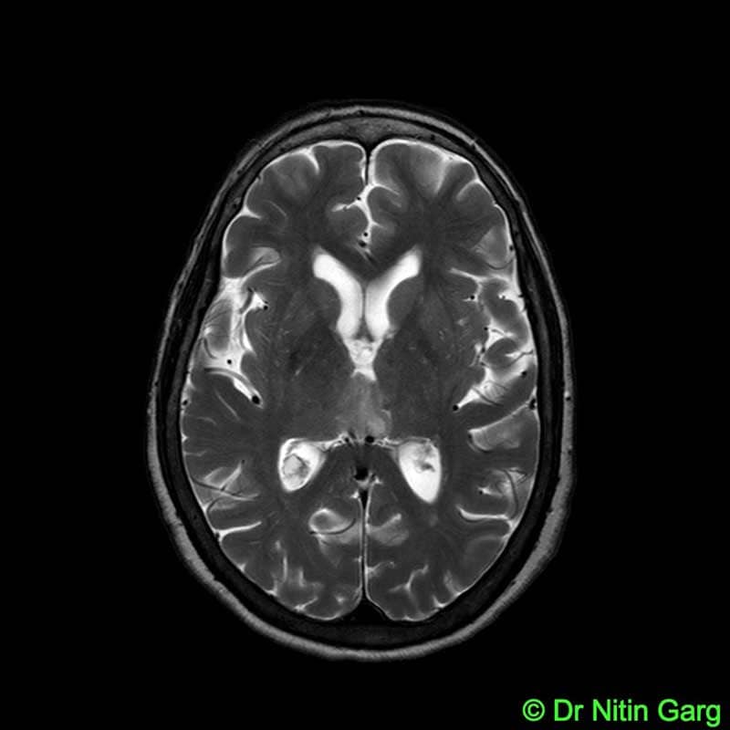
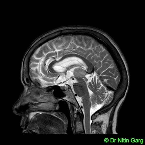
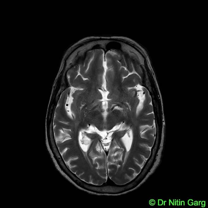
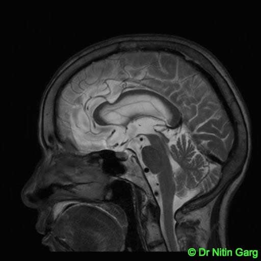
A 65 year old lady presented with headache, vomitting and drowsiness. MRI was suggestive of obstructive hydrocephalus due to a posterior third ventricular tumor (Figure 1). She underwent Endoscopic third ventriculostomy followed by biopsy from the same single burrhole. Patient was relieved of the symptoms and histopathology was suggestive of glioma. She was kept on follow up and 3 month MRI showed increase in the size of the glioma. Hence, adjuvant radiotherapy was considered. At 2 year follow up, the patient is doing well with no symptoms. Her MRI shows no residual tumor and resolution of hydrocephalus (Figure 2).
“Navigation” is a very useful in planning the approapriate trajectory for both ventriculostomy and biopsy and then planning the optimal position of the burrhole. The trajectory for ETV is more vertical and for biopsy from posterior third ventricle is more horizontal. Navigation helps to mark them on the workstation pre-operatively and then plan and mark the position of the burrhole somewhere in between.
Aids used: Endoscopy, Navigation.


