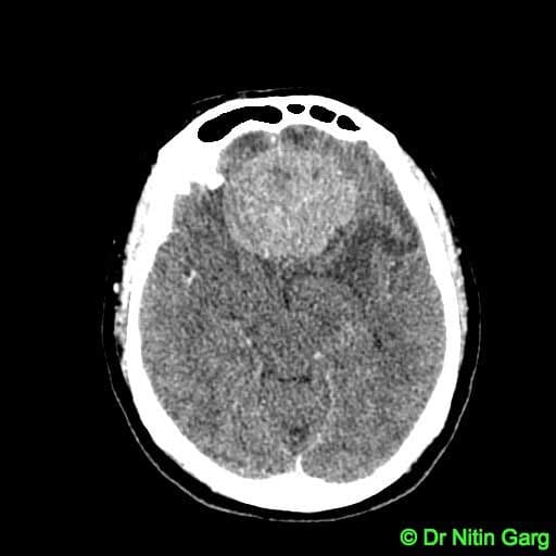
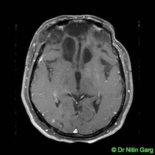
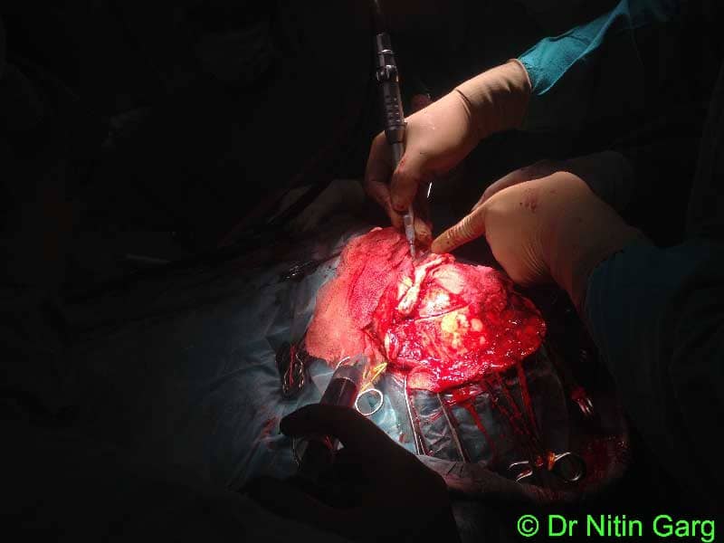
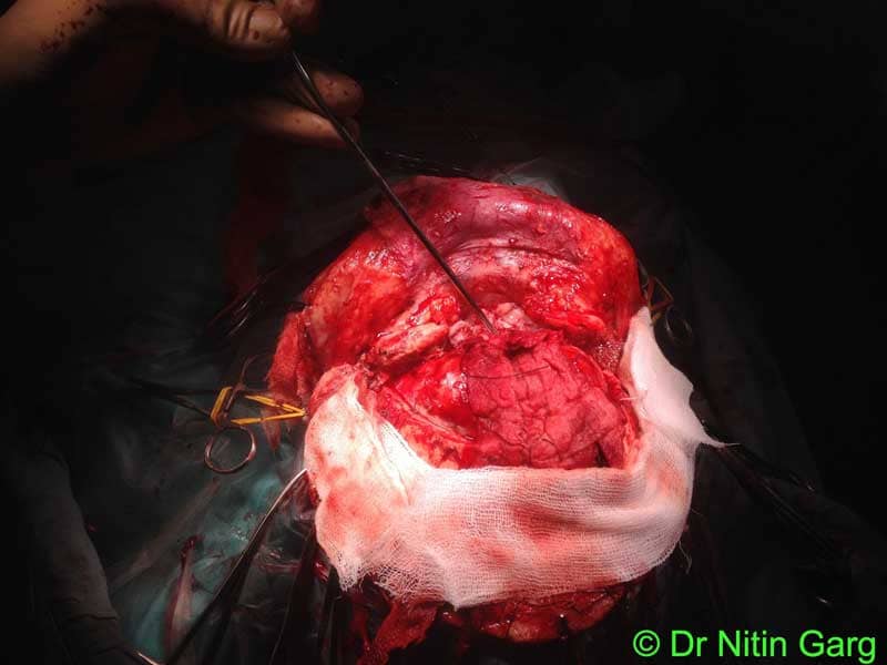
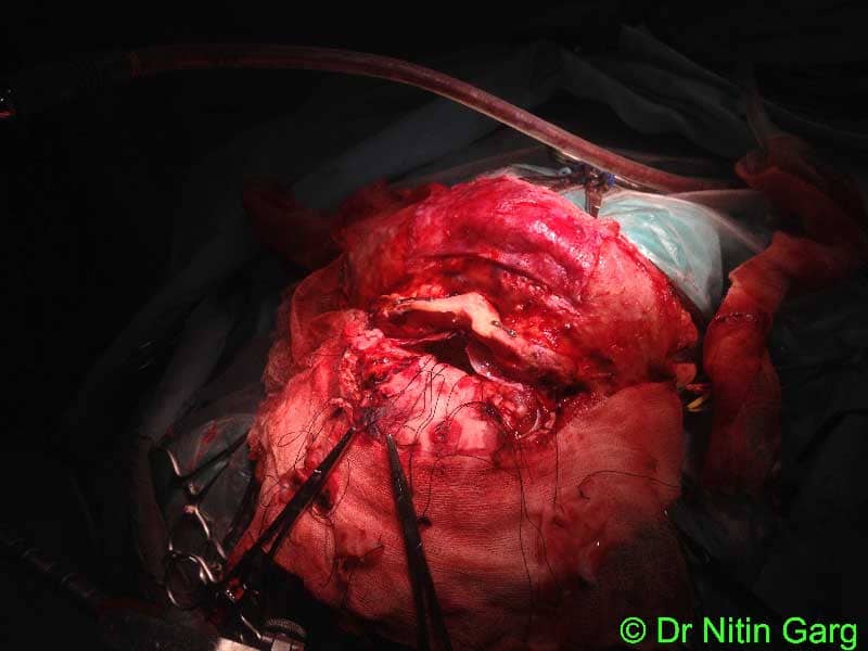
55 year old female patient presented with progressive visual deterioration. MRI of the brain showed a large suprasellar mass lesion suggestive of meningioma. She underwent bifrontal craniotomy, preglabellar approach and tumor excision. Intra-operatively, both the optic nerves were significantly stretched and pushed superiorly. The nerves appeared paler due to chronic compression. Internal tumor decompression was done, capsule gradually mobilised from both optic nerves and anterior communicating arteries. Thin layer of tumor adherent to the diaphragm sella was left behind. Both the optic foramen were unroofed to further decompress the optic nerves. Post operatively, patient made uneventful recovery. Over 3 months, there has been some improvement in vision. Follow up MRI shows adequate excision of the suprasellar mass with good decompression of the optic nerves and chiasma. There is thin enhancing tissue along the diaphragma sella. She has been planned for Gamma Knife radiosurgery for this residual tumor.
Aids used: Microscope, Neurosurgical drill with reciprocating saw attachment, Navigation, CUSA