Epilepsy is one of the most disabling neurological disorders. Amongst the various causes, tumor induced is one of the few surgically treatable causes. These tumors can be gliomas, meningiomas, metastasis commonly and few other histopathological types. DNET is one such rare tumor type . These are slow growing, benign tumors.
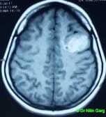
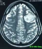
A 19 year old female patient presented with recurrent seizures. She had no features of rasied intracranial pressure. MRI showed a left sided well defined lesion with thinning of overlying bone. She underwent awake craniotomy and gross total excision of the tumor. Intra-operatively, the inner table of the bone was thinned out focally. The tumor was surfacing. Surface coagulated, internal decompression done. The tumor had a well defined plane from surrounding brain. Gross total excision achieved under constant clinical neurological monitoring. Patient tolerated the procedure well and recovered uneventfully and was discharged on the 2nd post-operative day.
Her histopathology was suggestive of DNET.
At one year follow up, the patient is completely off anti-epileptics and entirely seizure free.
Aids used : Microscope, CUSA, Awake craniotomy, navigation
“
Awake craniotomy” is a method of performing brain surgery while the patient is completely conscious. The aim is to monitor the neurological function continuously during the surgical procedure. This method is used in those patients who have a tumor located near epoquent areas of brain (like motor cortex, sensory cortex, speech areas) so that during removal of the tumor, these areas are not injured. The anaesthetist monitors the clinical condition continuously while the surgeon is removing the tumor. Whenever there is a slight impairment in the function being monitored (like hemiparesis or speech arrest), it signifies that the eloquent area is in proximity and further dissection is stopped immediately. In this manner, maximal safe resection of the tumor can be achieved without injury to the important area of the brain.