Cervical spondylotic myelopathy is a very common clinical condition managed by spine surgeons. Various approaches are used. Both ventral and dorsal methods are available for these patients.
In cervical sponylotic myelopathy, ventral cord compression is primary factor and ventral surgical methods can be used for upto 4 level disc prolapse. These include discectomy for a single to 2 level disc prolapse to corpectomy (upto 3 level corpectomy). Fusion is achieved by autologous bone graft and various titanium and PEEK prosthesis. Ventral methods require longer operating times, and patient should be in better general physical condition. Titanium constructs using plate and screws are used for fixation under C-arm guidance.
Dorsal appraoches include laminectomy (stand alone or with lateral mass fixation) and laminoplasty. These are more feasible and suited for patients with multiple level compression (more than 4), multiple co-morbid conditions, poor general physical condition. Posterior approaches result in indirect decompression.
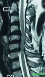
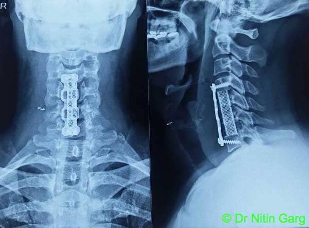
Case: a 40 year old patient presented with progressive wakness of all four limbs of 2 months duration. MRI (Figure 1) showed 3 level disc prolapse with cord compression. CT scan (Figure 2) revealed multiple posterior osteophytes. He underwent anterior approach, 2 level corpectomy, 3 level discectomy and fusion with titanium cage filled with bone autograft and titanium plates and screws (Figure 3)
Aids used : Microscope, Neurosurgical drill, C-arm.
“High speed neurosurgical drills” aid in corpectomy and removal of posterior osteophytes. These drills have small diameter bits (2.4-4mm) which core out the bone till a very thin layer of cortex is left behind. This is then removed with a curette or 1mm bone punches. Such method minimises the risk of injury to the cord.
“Microscope” is essential to achieve the adequate and safe neural decompression.
“C-arm” is useful at multiple steps. In the beginning, it is used to mark the incision according to the level of decompression planned. Subsequently, once prevertebral space is reached, the level of interest is reconfirmed. Subsequently, appropriate position and size of the cage and plate and screw trjectory is all done under C-arm guidance.